Mechanical Imaging of breast was the first project of Artann, funded by NIH in 1995. Physical basis and general principles and algorithms underlying the breast MI technology were disclosed in the USA patent filed by Sarvazyan in 1992. The Breast Mechanical Imager (BMI) developed under several NIH SBIR grants electronically captures the sense of touch to provide a sensitive, repeatable, quantitative, and permanently stored record of the breast examination. The device includes a probe with a pressure sensor array, an electronic unit, and a laptop computer. The Breast Mechanical Imager, cleared by FDA for breast lesion visualization and documentation, is currently on the market produced by MTI (Los Angeles, CA) under trade name SureTouchTM.
Clinical data have demonstrated BMI‘s capability for characterizing and differentiating benign and malignant breast lesions. Histologically confirmed malignant breast lesions demonstrated increased hardness and strain hardening as well as decreased mobility in comparison with benign lesions. Statistical analysis of data on 147 benign and 32 malignant lesions revealed BMI’s average sensitivity of 91.4% and specificity of 86.8% with a standard deviation of ±6.1%. The study had indicated BMI potential for a cost effective device for cancer diagnostics that could be positioned as an adjunct to mammography and utilized as a screening device for breast cancer detections. The table below shows a few examples of clinical data obtained by BMI.
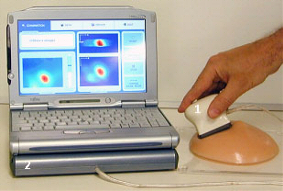
General view of Breast Mechanical Imager. The device comprises a probe with 2-D pressure sensor array (1), an electronic unit (2), and a laptop computer with touch screen capability
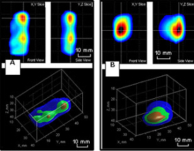
5Processed data for two clinical cases demonstrate the ability of MI technology to produce 3-D images of lesions. A - two cysts; B - invasive ductal carcinoma
Advantages of BMI include inherently low cost, ease-of-use, portability, and minimal training required. Cost analysis of currently used breast screening and diagnostic modalities showed that BMI may be over ten times more cost-effective than mammography.
Selected Publications and Patents
- Egorov V, Kearney T, Pollak SB, Rohatgi C, Sarvazyan N, Airapetian S, Browning S, Sarvazyan A. Differentiation of benign and malignant breast lesions by mechanical imaging. Breast Cancer Res Treat 2009; 11:118(1):67-80.
- Egorov V, Sarvazyan AP: Mechanical Imaging of the Breast. IEEE Transactions on Medical Imaging 2008; 27(9):1275-87.
- Sarvazyan A, Egorov V, Son JS, Kaufman CS: Cost-effective screening for breast cancer worldwide: current state and future directions. Breast Cancer: Basic and Clinical Research 2008; 1:91–9.
- Pashko DA, Pyt’ev YP, Sarvazyan AP, Skovoroda AR: On the ultimate potentialities of classification of pathologies in the problem of diagnostics of breast cancer. Pattern Recognition and Image Analysis 1996; 6: 510-22.
- Pashko DA, Pyt’ev YP, Sarvazyan AP: Minimax evaluation of parameters of a nodule in diagnosing breast cancer with the use of a force sensor array. Bulletin of Moscow State University 1996; Ser 3 Physics Astronomy:18-25.
- Sarvazyan A, Egorov V. Real time mechanical imaging of the prostate. USA Patent 6,569,108, May 27, 2003.
- Sarvazyan A, Egorov V. Self-palpation device for examination of breast with 3-D positioning system. USA Patent 6,595,933, July 22, 2003.
- Sarvazyan A, Egorov V. Apparatus and method for mechanical imaging of breast. USA Patent 6,620,115, Sep 16, 2003.
- Sarvazyan AP, Egorov V. Self-palpation device for examination of breast. USA Patent 6,468,231, Oct 22, 2002.
- Sarvazyan A. Method and device for mechanical imaging of breast. USA Patent 5,860,934, Jan 19, 1999.
- Sarvazyan A. Device for breast haptic examination. USA Patent 5,833,633, Nov. 10, 1998.